Basal Cell Carcinoma (BCC) is the most common type of skin cancer in Australia, largely due to chronic sun exposure. Although it rarely spreads to distant parts of the body, BCC can cause significant local tissue damage if left untreated.
Because these cancers tend to grow slowly and remain confined to the skin, complete surgical removal is the most effective form of treatment. As with any skin cancer procedure - the goal is to remove the cancer completely while preserving the function and appearance of the affected area.
Excision of a BCC from cosmetically sensitive areas such as the nose, eyelids, ears, or lips is particularly challenging and should be performed by a surgeon with substantial experience. Dr Schaefer performs multiple BCC excisions every week at Brisbane Private Hospital.
Basal cell carcinomas can be diagnosed by a trained GP or dermatologist. In some cases, a biopsy is required for diagnosis which identifies the cancer type and subtype. These findings, along with the lesion’s size, depth, and location, guide Dr Schaefer’s surgical approach.
There are several forms of BCC:
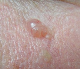
Nodular BCC:
A pearly, raised lump with visible blood vessels; usually well defined.
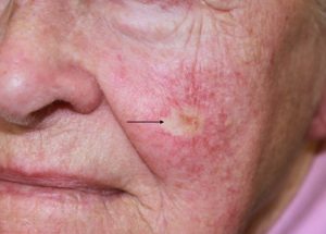
Superficial BCC:
Thin, red or scaly patches that spread across the skin surface.

Infiltrative (or morphoeic) BCC:
Scar-like and often ill-defined, extending beyond visible borders.
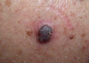
Pigmented BCC:
A rare variant containing melanin, giving it a brown, blue, or black colour that can resemble melanoma, but with the same localised growth pattern as other BCC types.
Fibrosing and infiltrative BCCs are particularly challenging to fully remove, as they can send microscopic extensions under the skin. In these cases, Dr Schaefer often takes wider margins or may recommend Mohs micrographic surgery to ensure all cancer cells are removed.
BCC removal can be performed under local anaesthesia in his rooms on Wickham Terrace, or under a general anaesthetic at Brisbane Private Hospital. There are many factors which influence this decision including the type of skin cancer, the location of the skin cancer and patient preference.
During surgery, Dr Schaefer carefully removes the skin cancer along with a small margin of healthy tissue surrounding the skin cancer. The recommended margin typically ranges from 2 mm to 5 mm, depending on the subtype of the BCC.
The specimen is precisely marked and sent to a pathologist to confirm whether the cancer has been fully removed.
Once the lesion has been excised, Dr Schaefer focuses on achieving a repair that restores both function and appearance. The reconstruction method depends on the site, size, and depth of the excision.
Common approaches include:
Pathology results are available within the week after surgery. If all margins are clear, no further surgery is required. If a margin is closer than desired, a re-excision procedure may be recommended to ensure the cancer is fully removed.
After BCC removal surgery, the wound is dressed and patients receive written and verbal instructions on care, pain relief, and activity restrictions. Stitches are removed within one week of surgery.
Healing typically takes one to two weeks, during which time strenuous activity and swimming should be avoided. Most patients return to normal routines soon after surgery.
All surgeries carry risks, and Basal Cell Carcinoma excision is no exception. Possible complications include:
Once a BCC has been removed, the risk of developing another remains higher than average. Ongoing skin checks every 6–12 months are strongly advised.