Melanoma is the most serious type of skin cancer, capable of spreading (metastasising) if not treated early. Early surgical excision with appropriate margins offers the best chance of cure. Dr Schaefer regularly performs melanoma removal procedures at Brisbane Private Hospital, located in Spring Hill.
After the initial biopsy confirms melanoma, the Breslow thickness (depth in millimetres) and other histological features guide what margin of healthy skin tissue must be removed.
Dr Schaefer will analyse your individual pathology report and plan a removal margin that balances oncologic safety with skin preservation whilst always following evidence-based guidelines
On the day of surgery (often as a day-case, under local or general anaesthesia depending on size/location):
Once the melanoma is removed, attention turns to wound closure. Dr Schaefer selects the approach best suited to your site and size of removal:
Direct closure / primary closure: for small lesions, the wound edges can generally be stitched directly together. 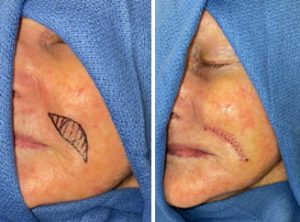
Skin grafting: a patch of skin (often from a nearby area) transplanted over the excision site.
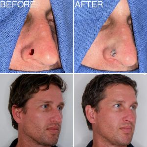
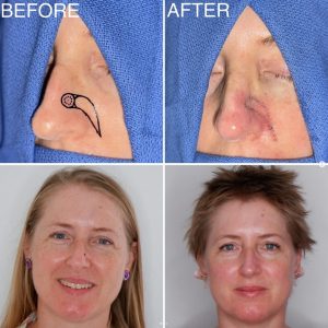
Local flap repair: nearby tissue is rotated or shifted to cover the defect, usually with good colour and texture match.
Combined or staged reconstructions: for more complex defects, reconstruction may occur in phases for optimal result.
Dr Schaefer’s reconstructive choices are always driven by the goal of restoring appearance and function while minimising visible scarring.
The excised tissue is examined under a microscope. If margins are confirmed clear, your melanoma has been fully removed. If any margin is involved (cancer cells extend to the edge), a further excision may be required.
Follow-up includes regular skin checks, monitoring for recurrence, and education on sun protection and skin surveillance.
All surgeries carry risks, and melanoma excision is no exception. Possible complications include: